Archive : Article / Volume 2, Issue 2
- Case Report | DOI:
- https://doi.org/10.58489/2836-5828/007
Leukemia Infiltration Secondary Acute Renal Damage
¹MD, Resident, Department of Internal Medicine, Ankara City Hospital, Ankara, Turkey
2MD, Physician, Division of Nephrology, Ankara City Hospital, Ankara, Turkey
3MD, Physician, Department of Pathology, Ankara City Hospital, Turkey
4MD, Associate Professor, Department of Nephrology, Ankara City Hospital, Ankara, Turkey
5MD, Professor, Department of Nephrology, Ankara City Hospital, Ankara, Turkey
Amed Trak, MD, Resident, Department of Internal Medicine, Ankara City Hospital, Ankara, Turkey
Amed Trak, et.al. Leukemia Infiltration Secondary Acute Renal Damage. Arch. Urol. Nephrol. (2023). Vol. 2, Iss. 2. DOI: 10.58489/2836- 5828/007
© 2023 Amed Trak, this is an open-access article distributed under the Creative Commons Attribution License, which permits unrestricted use, distribution, and reproduction in any medium, provided the original work is properly cited.
- Received Date: 27-02-2023
- Accepted Date: 24-03-2023
- Published Date: 03-04-2023
Chronic lymphocytic leukemia, Leukemic infiltration, Acute renal damage
Abstract
Chronic lymphocytic leukemia is a disease that it is developed by cumulation of mature-looking small lymphocytes in blood, bone marrow, lymph nodes,s pleen and other lymphatic organs and tissues.It can be attained as asymptomatic lymphocytosis in old patients;on the other hand, kidney dysfunction can also be seen as a preliminary finding. Leukemia infiltration secondary acute renal damage can rarely be seen while renal damage is being occured because of many different reasons in chronic lymphocytic leukemia patients. Here, we aimed to review the literature by presenting 70-year-old patient who was under hematology follow-up due to chronic lymphocytic leukemia, who was hospitalized in our clinic because of acute kidney damage in routine examinations and was found to have leukemic infiltration in pathology.
Introduction
Chronic lymphocytic leukemia(CLL) is a disease that it is developed by cumulation of mature-looking small lymphocytes in blood,bone marrow,lymph nodes,spleen and other lymphatic organs and tissues [1]. It is known that chronic lymphocytic leukemia(CLL) can be accompanied by renal failure.Kidney failure incidence was found %7,5 in cohort study which was conducted by Mayo Clinic with more than 2000 CLL patients [2]. CLL’s renal damage reasons can be listed as ureteral obstruction caused by lymphadenopathy pressure,tumor lysis syndrome(TLS) which can be developed after treatment and direct kidney infiltration which is rarely being seen.Pathologies like light chain nephropathy,renal amyloidosis, membranoproliferative glomerulonephritis(MPGN), granulomatous interstitial nephrite and minimal change disease were reported among other uncommon reasons [2]. Here;in a patient with CLL,our aim was to present a case with lymphoid cell infiltration in kidney biopsy performed due to Acute Kidney Injury (AKI).
Case Presentation
A 70-year-old male patient with a diagnosis of hypertension (HT), dementia, chronic obstructive lung disease, and stage 1 CLL for 1 year, with creatinine of 1.1 mg / dL a month earlier and a creatinine value of 1.76 mg / dL in routine control, was hospitalized to our service with the diagnosis of AKI. It was learned that he was followed up in the Hematology Clinic due to stage 1 CLL and he was under conservative follow-up, because immunosuppressive therapy was not seen as a necessity. The patient having had no active complaint in examination for AKI etiology did not have nephrotoxic agent usage history;besides,prerenal and postrenal reasons were also excluded.In the physical examination,blood pressure arterial was 140/90 mmHg and cardiovascular,respiratory system and abdominal examination was normal. A diameter of 1.5 cm in the right axillary area and a diameter of 2 cm palpable lymphadenopathy in the right anterocervical area were pathologically identified. In the patient’s laboratory values; urea: 52 mg / dL (<49 mg / dL), creatinine: 2.1 mg / dL (0.5-1.1 mg / dL), sodium: 144 mEq / L (137-2-146 mEq / L), potassium: 5.03 mEq / L (3.5-5.5 mEq / L), WBC: 25.900 / mm3 (3.9-10.2 / mm3), neutrophil: 2100 / mm3 (1.5-7.7 / mm3), lymphocyte: 18.300 / mm3 (1.1-4 , 5 / mm3) hemoglobin: 11.6 g / L (12-15.6 g / dL), thrombocyte: 193.000 / mm3 (150.000-370.000 / mm3) and mature lymphocytes and basket cells compatible with lymphocytosis were seen in peripheral smear. It was reported that right kidney dimensions were 128 × 55 mm, parenchymal thickness was 14 mm and echogenicity strength increased grade 1, left kidney dimensions were 130 × 50 mm, parenchymal thickness was 16 mm and echogenicity strength increased grade 1 in the urinary system ultrasonography (USG) of the patient. Complement levels for AKI etiology were in normal limits and ANA, Anti-dsDNA, p-ANCA, c-ANCA, anti-GBM were detected negative. During the follow-ups,the patient’s creatinin values progressively increased up to 2.7 mg/dL;that is why,3 core renal biopsies were taken with 16G tru-cut needle accompanied by USG in terms of AKI etiology 3 of 14 glomeruli were seen as global sclerotic in the pathological examination of renal biopsy.Light periglomerular fibrosis and retraction in capillary glomus were tracked in the other glomeruli. There were condense lymphocytic infiltration like a patch in the interstitium and infiltrative cells were monitored in a monotonous character.Expansion of glomeruli and increased focal thickness in the basal membranes,dilatation in some of the tubules and flattening of the tubular epithelial cells were observed.In the immunohistochemical examination; CD5,CD23 and diffuse positive staining with repeatedly done CD20 were detected;moreover, positiveness was observed in a limited number of lymphoid cells with CD3(figures 1,2 and 3). CLL disease was interpreted for benefit of kidney involvement when the immunohistochemical symptoms were evaluated with the clinical history of the case. The patient,whose creatinine regressed spontaneously up to 1.6 mg/dL during his follow-ups, was directed to the Hematology Clinic for the treatment of the primary disease and was discharged.The patient,who has been on our polyclinic follow-up for 6 months, had no chemotherapy indication in terms of hematological aspect because of his being phase 1 and was taken to the conservative follow-up.His creatinine values got stuck in 1.5 mg/dL during the follow-ups and it was accepted as chronic kidney disease( CKD).
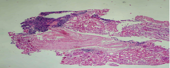
Figure 1: Monotonous small lymphoid cells infiltration in kidney needle biopsy material (H&E x40)
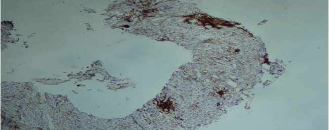
Figure 2: CD5 IHC stain is strongly positive small lymphocytes (×40)
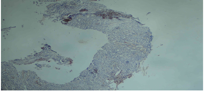
Figure 3: CD23 IHC stain is positive in small lymphocytes (×400)
Discussıon
hronic lymphocytic leukemia is a lymphoid malignancy characterized with the cumulation and increasing of the mature CD5 + B cells in blood,bone marrow and lymphoid tissues [1]. The average age in CLL is 71. The incidence frequency of the disease in the USA is 4.5/100.000. Male and female incidence rate is 2:1. The incidences get closer to each other as people grow older. They are seen at the equal rate in both sexes even at the ages over 80 [3]. CLL diagnosis is made by the presence of over 5.000/μL mature B lymphocytes in peripheral blood at least for 3 months. .[4] For CLL; CD5, CD19 and CD23 are found in characteristic immunophenotype whereas CD20, surface immunoglobulin, CD79b and FMC7 are barely found [4]. If lymphocytes are positive with CD5,two possibilities are extremely high: CLL or mantle cell lymphoma(MCL).CD23 is generally positive in CLL cells;besides,it is possible to diagnose correctly because CD23 is negative in MCL.CLL renal involvement has been confirmed in our case by observing diffuse CD5, CD20 and CD23 staining in lymphoid cells.
In CLL which shows a highly variable clinical course; after the diagnosis, some of the patients are extremely silent and asymptomatic and live for many years without progressing, while some of them tend to progress rapidly although they are either advanced or early phase at the time of diagnosis [5]. In the patients who has been started to the treatment; the most common signs and symptoms are fever,night sweating,weight loss,organomegaly, anemia associated with bone marrow failure and thrombocytopenia [2]. In CLL; AKI associated with infiltration is a very rarely seen complication despite the fact that it is reported at %60-90 in renal involvement autopsy series. AKI can be detected as a first sign of CLL in some cases.It is presumed that leukemic infiltration causes a tubular obstruction even though kidney damage mechanism is not exactly known [6]. Except that,CLL also causes gromerular pathologies.MPGN (%36) and membranous nephropathy (%18) are the mostly encountered gromerular pathologies [2]. The other rarely seen glomerulonephritis types are minimal change disease and focal segmental glomerulosclerosis.Tumor lysis syndrome in CLL is scarcely seen but it is thought that venetoclax,which is bcl-2 inhibitor and used in the treatments of %6 of the patients,causes it [2].
Another complication of CLL is amyloidosis. 18 (%55) of the patients are diagnosed with AL amyloidosis with renal biopsy in a study including 33 CLL patients who are taken place in the literature. While plasma cell clones brought about amyloidosis in 14 of these patients,it was identfied that CLL clones caused amyloidosis in the other 4 patients [7]. Clinical staging system was based on defining prognosis in CLL;furthermore,only physical examination and peripheral smear are needed in determining clinical staging [10]. In the notifications published between 1966 and 1973, Rai et al. developed a CLL staging system that could prospectively differentiate patients based on their overall appearance for survival [2]. After that, a new classification was introduced by Binet et al. There have been some deficiencies although there is a meaninful relationship between stage and survival in both classifications.Modified Rai and Binet classification is stated in table 1 [5]. Patients with intermediate (Stage I and II) and high risk (Stage III and IV) according to the modified Rai classification or stage B and C according to the Binet classification generally benefit from treatment. Some of these patients (Rai intermediate risk and Binet stage B ) can be followed without treatment unless any progression symptoms or signs appear [5].
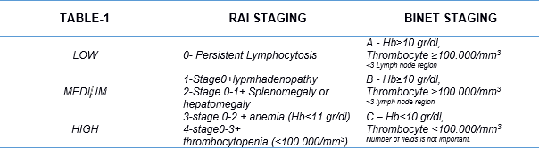
Table.1: Modified Rai and Binet Staging System
AKI associated with leukemic infiltration was described in previously reported cases in the literature. Renal involvement couldn’t have been related to the staging in CLL patients.Renal involvement was shown by doing biopsy and different chemotherapy regimens were applied to the cases involved in the literature. Kidney function recovery was observed as a result of the treatment and recovery of kidney functions via regression was also observed in some cases [2].
As a result; leukemic infiltration should always be kept in mind when CLL patients apply with the kidney dysfunction. The case we have presented is stage 1 CLL and there is no treatment indication in terms of hematology according to current modified Rai and Binet classifications.It has been observed that renal funtions also recover as a consequence of the treatment of primary disease with chemotherapy in AKI caused by renal infiltration in the literature.On the other hand; chemotheraphy couldn’t have been given because of his being stage 1 in our case.It was observed that his creatinine value stopped at 1.5 mg/dL by the end of the 6th month on our polyclinical follow-ups. On account of this; we are agreed that along with liver and spleen involvement in staging of CLL, lymphocytic infiltration proven by renal biopsy to be performed in the presence of accompanying renal dysfunction should be included in the classification, and CLL staging and chemotherapy indication should be re-evaluated, and cases accompanied by AKI should be given a chance to treat.
References
- Delgado, Julio, Ferran Nadeu, Dolors Colomer, and Elias Campo. “Chronic lymphocytic leukemia: from molecular pathogenesis to novel therapeutic strategies.” Haematologica 105, no. 9 (2020): 2205.
- Wanchoo R , Ramirez C.B., Barrientos J , Jhaveri K. et al “Renal Involvement in Chronic Lymphocytic Leukemia” Clin Kidney J 2018 Oct;11(5):670-680.
- Jennifer A. Woyach, John C. Byrd Harrison, E. Braunwald, A. S. Fauci, D. L. Kasper et al, “Principles of Internal Medicine “ 1024–1034, McGraw-Hill, North America, 2018.
- Hallek, Michael, Bruce D. Cheson, Daniel Catovsky, Federico Caligaris-Cappio, Guillaume Dighiero, Hartmut Döhner, Peter Hillmen et al. “Guidelines for the diagnosis and treatment of chronic lymphocytic leukemia: a report from the International Workshop on Chronic Lymphocytic Leukemia updating the National Cancer Institute–Working Group 1996 guidelines.” Blood, The Journal of the American Society of Hematology 111, no. 12 (2008): 5446-5456.
- Türkiye Hematoloji Derneği Ulusal Tedavi ve Tanı Klavuzu 2012 syf:3,6
- Ferreira, Ana Carina, Sandra Brum, Dulce Carvalho, Isabel Pataca, Fernanda Carvalho, Maria Ceu Santos, and Joao Ribeiro Santos. “Renal dysfunction due to leukemic infiltration of kidneys in a case of chronic lymphocytic leukemia.” Hemodialysis International 14, no. 1 (2010): 87-90.
- Kourelis, Taxiarchis V., Morie Gertz, Clive Zent, Martha Lacy, Robert Kyle, Prashant Kapoor, Steven Zeldenrust et al. “Systemic amyloidosis associated with chronic lymphocytic leukemia/ small lymphocytic lymphoma.” American journal of hematology 88, no. 5 (2013): 375-378.
- Demir, Vahit, Selda Kahraman, A. Katgı, Özden Pişkin, Güner Hayri Özsan, Fatih Demirkan, Bülent Ündar, and Mehmet Ali Özcan. “Kronik lenfositik lösemi hastalarının genel klinik değerlendirilmesi.” Dokuz Eylül Üniversitesi Tıp Fakültesi Dergisi 26, no. 1 (2012): 9-19.
- Hewamana, Saman, Chris Pepper, Chris Jenkins, and Clare Rowntree. “Acute renal failure as the presenting feature of leukaemic infiltration in chronic lymphocytic leukaemia.” Clinical and experimental nephrology 13, no. 2 (2009): 179-181.
- Çağlıyan, Gülsüm Akgün, Nilüfer Aslankarasoy, and Oktay Bilgir. “Kronik Lenfositik Lösemili Hastaların Değerlendirilmesi: Tek Merkez Deneyimi 1.” 43-48.


