Archive : Article / Volume 2, Issue 1
- Research Article | DOI:
- https://doi.org/10.58489/2836-5003/009
Morphological Changes in Neurons of The Parietal Cortex and Hippocampus of Rats with Partial Cerebral Ischemia
- Grodno State Medical University, 80, Gorkogo St., 230009, Grodno, Republic of Belarus
E.I. Bon
Bon E.I., Maksimovich N.Ye., Zimatkin S.M., Lelis A.R., (2023). Morphological Changes in Neurons of The Parietal Cortex and Hippocampus of Rats with Partial Cerebral Ischemia. Archives of Immunology Research and Therapy. 2(1). DOI: 10.58489/2836-5003/009.
© 2023 Bon E.I, this is an open access article distributed under the Creative Commons Attribution License, which permits unrestricted use, distribution, and reproduction in any medium, provided the original work is properly cited.
- Received Date: 31-01-2023
- Accepted Date: 04-02-2023
- Published Date: 06-02-2023
neurons; parietal cortex; hippocampus; rats; cerebral ischemia
Abstract
The study of the brain in health and disease is a relevant and promising area of modern science and, in this regard, a frequent topic of dissertation research. Cerebral ischemia leads to a number of general and local metabolic and functional disorders, the pathogenesis of which is complex, many-sided and largely unclear. The absence of pronounced morphological changes in the simulation of PCI after 1 hour in rats is explained by the compensation of blood circulation in the circle of Willis.
Introduction
The study of the brain in health and disease is a relevant and promising area of modern science and, in this regard, a frequent topic of dissertation research. Cerebral ischemia leads to a number of general and local metabolic and functional disorders, the pathogenesis of which is complex, many-sided and largely unclear [2, 4-10].
Purpose - changes in the morphology of neurons in the parietal cortex and hippocampus of rats with partial cerebral ischemia.
Methods
The experiments were carried out on 24 male outbred white rats weighing 260±20 g in compliance with the requirements of the Directive of the European Parliament and of the Council № 2010/63/EU dated 22.09.2010 on the protection of animals used for scientific purposes.
The choice of experimental animals is due to the similarity of the angioarchitectonics of the brain of rats and humans. Modeling of cerebral ischemia (CI) was performed under conditions of intravenous thiopental anesthesia (40-50 mg/kg).
The studies used models of partial (PCI), cerebral ischemia.
Partial CI (PCI) was modeled by ligation of one common carotid artery (CCA).
A morphological study of the size and shape of neuronal perikaryons, determination of the number of neurons with different degrees of cytoplasmic chromatophilia was carried out [Bon L.I., 2019, Danilov R.K., 2011].
Statistical data processing
To prevent a systematic measurement error, brain samples from the compared control and experimental animals were treated under the same conditions.
As a result, of morphometric and cytophotometric studies, quantitative continuous data were obtained, which were processed using the licensed computer program Statistica 10.0 for Windows (StatSoft, Inc., USA).
Since the experiment used small samples that had an abnormal distribution, the analysis was carried out using nonparametric statistics methods. The data are presented as Me (LQ;UQ), where Me is the median, LQ is the value of the lower quartile; UQ is the value of the upper quartile. Differences between groups were considered significant at p<0>
Results
The study of morphological changes in partial CI was carried out in the parietal cortex and hippocampus of rats.
For this purpose, a histological study was carried out, for which, after decapitation of the animal, the brain was quickly removed, pieces of the anterior part of the cerebral cortex were fixed in Carnoy's fluid. Localization of the parietal cortex and hippocampus cortex in histological preparations of the rat brain was determined using a stereotaxic atlas.
Serial paraffin sections were stained with 0.1% toluidine blue by the Nissl method and for the detection of ribonucleoproteins by Einarson.
The study of histological preparations, their microphotography, morphometry and densitometry of the chromogen sediment using an Axioscop 2 plus microscope (Zeiss, Germany), a digital video camera (LeicaDFC 320, Germany) and ImageWarp image analysis program (Bitflow, USA).
At least 30 neurons of the fifth layer of the parietal cortex and the pyramidal layer of the field CA1 of the hippocampus were evaluated in each animal, which provided a sufficient sample size for subsequent analysis.
To assess the severity of ischemic damage to the cerebral cortex under CI, we studied changes in the size and shape of neuronal perikaryas, as well as the degree of staining of their cytoplasm (chromatophilia).
The area of neurons (S) and the elongation of their bodies indicate the state of the cytoskeleton and water and electrolyte balance, which can be disturbed during cerebral ischemia. Chromatophilia of the cytoplasm, in turn, reflects the functional activity of the neuron [2, 6].
The study of changes in the size and shape of neuronal perikarya was carried out by assessing their area, form factor (4πS / P2 - index of sphericity and folding), elongation factor - index of sphericity (Dmax/Dmin) using the ImageWarp image analysis program (Bitflow, USA).
In histological preparations, the number of large pyramidal neurons per unit area of sections of the cerebral cortex was determined. Among the total number of neurons, cells were distinguished according to the intensity of cytoplasm staining (chromatophilia): normochromic - moderately stained; hyperchromic - dark; hyperchromic wrinkled - very dark, with deformed perikarya; hypochromic - pale colored; shadow cells are unstained, with vacuolated nuclei. The number of each cell type was counted [6, 8].
With partial cerebral ischemia, no statistically significant differences were found between the parameters characterizing the size and shape of neuronal perikarya, the number of hyperchromic shriveled neurons and shadow cells in the PCI group, compared with the control, both in the parietal cortex and in the hippocampus (table 1, figure 1), except for a smaller number of normochromic (p<0>
Table 1 - Sizes and shape of perikaryons of neurons in the parietal cortex and hippocampus of rats with partial cerebral ischemia (PCI) and the control group, Me (LQ; UQ)
Animal groups | Areas of the cerebral cortex | |
parietal cortex | hippocampus | |
area, mcm2 | ||
Control | 144,6(130,5;153,8) | 108,6(99,7;122,3) |
PCI | 161(143;165) | 96(92;100) |
elongation factor, units | ||
Control | 1,2(1,1;1,2) | 1,2(1,2;1,2) |
PCI | 1,2(1,1;1,3) | 1,1(1,1;1,2) |
form factor, unit | ||
Control | 0,9(0,9;0,9) | 0,9(0,9;0,9) |
PCI | 0,9(0,8;0,9) | 0,9(0,8;0,9) |
Note: PCI - partial cerebral ischemia
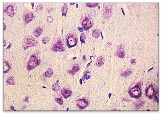
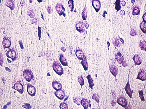
Figure 1. Neurons of the fifth layer of the parietal cortex in rats with partial cerebral ischemia (PCI). A - control (the predominance of normochromic neurons). B - PCI (the predominance of hyperchromic neurons). Digital micrograph. Nissl coloring. SW. lens x 40
Table 2 - The number of different forms of neurons according to the degree of chromatophilia of the cytoplasm of the parietal cortex and hippocampus of rats with partial cerebral ischemia (PCI) and the control group, Me (LQ; UQ)
Animal groups | Areas of the cerebral cortex | |
parietal cortex | hippocampus | |
normochromic neurons / mm2 | ||
Control | 3283(3216;3283) | 3216(3149;3283) |
PCI | 2678(2678,2678)* | 2712(2678,2745)* |
hyperchromic neurons / mm2 | ||
Control | 201(201;268) | 167,5(134;201) |
PCI | 569,5(536;670)* | 536(536;603)* |
hyperchromic shriveled neurons / mm2 | ||
Control | 134(67;134) | 33(0;134) |
PCI | 100,5(67;134) | 100,5(0;134) |
shadow-cells / mm2 | ||
Control | 134(0;134) | 0(0;134) |
PCI | 67(0;134) | 134(0;134) |
Note: * - p0.05 compared with the control group, PCI - partial cerebral ischemia
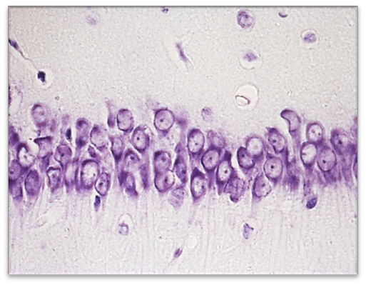
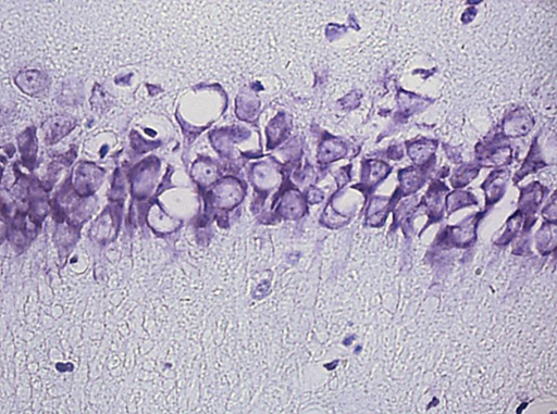
Figure 2. Neurons of the pyramidal layer of the field CA1 of the hippocampus of the brain of rats with partial cerebral ischemia (PCI). A - control (the predominance of normochromic neurons). B - PCI (the predominance of hyperchromic neurons). Digital micrograph. Nissl coloring. SW. lens x 40
Thus, the absence of pronounced morphological changes in the simulation of PCI after 1 hour in rats is explained by the compensation of blood circulation in the circle of Willis.
References
- Batin, N. V. (2008), Computer statistical data analysis: textbook. allowance / N. V. Batin. - Minsk: Institute of preparation. scientific personnel of the National Academy of Sciences of Belarus. - 160 p.
- Bon L.I. (2019), Changes in the chromatophily of the cytoplasm of large pyramidal neurons in the neocortex of the rat brain in postnatal ontogenesis / L.I. Bon, S.M. Zimatkin // Bulletin of the Smolensk State Medical Academy. - No. 1. - P. 10-16.
- Rebrova, O. Yu. (2003), Statistical analysis of medical data. Application of the application package Statistica / O. Yu. Rebrova. - M.: MediaSphere,. - 312 p.
- Guide to histology / edited by R.K. Danilov. (2011), - 2nd ed., corrected. and additional - St. Petersburg.:
- Bon, L.I. (2018), Morphofunctional disorders in the hippocampus of rats with subtotal ischemia / E.I. Bon, N.E. Maksimovich, S.M. Zimatkin // Bulletin of the Smolensk State Medical Academy. - No. 1. - S. 24-29.
- Zimatkin, M. (2017), Dark brain neurons / L.I. Bon, S.M. Zimatkin // Morphology. - No. 6. - P.81-86.
- Semchenko, V.V. (1999), Post-anoxic encephalopathy / V.V. Semchenko // Omsk. - 448 p.
- Bon, L.I. (2018), Effects of experimental cerebral ishemia on metabolic characteristics of parietal cortex neurons / L.I. Bon, N.Ye. Maksimovich, S.M. Zimatkin // Bioprocess Engineering. - Vol. 2(1). – P. 1-5.
- Clemens, J.A. (2000), Cerebral ischemia: gene activation, neuronal injury, and the protective role of antioxidants / J.A. Clemens // Free Radic. Biol. Med. - Vol. 28. - P. 1526-1531.
- Gallyas, F. (2009), Supravital microwave experiments support that the formation of


