
Case Report | DOI: https://doi.org/10.58489/2836-2225/011
1 MD, Obstetrics and Gynecology, Post-Graduate, Orotta College of Medicine and Health Science, Ministry of Health, Asmara, Eritrea.
2 MD, MPH, Obstetrician and Gynecologist, Associate Professor, Orotta College of Medicine and Health Science, Post-Graduate, Orotta National Referral Maternity Hospital, Ministry of Health, Asmara, Eritrea.
3 MD, Dekemhare Hospital, Zoba Debub, Ministry of Health, Eritrea.
*Corresponding Author: Berhe Tesfai
Citation: Berhe Tesfai, Okbu Frezgi, Saron Abraham, Malede Birara, and Hailemichael Gebremariam (2023), Huge Rare Vaginal Fibromatosis in Postmenopausal Woman, Case Report and Literature Review, 2022, International Journal of Reproductive Research 2(1): DOI: 10.58489/2836-2225/011
Copyright: © 2023 Berhe Tesfai. This is an open-access article distributed under the terms of The Creative Commons Attribution License, which permits unrestricted use, distribution, and reproduction in any medium, provided the original author and source are credited.
Received: 03 March 2023 | Accepted: 28 March 2023 | Published: 06 April 2023
Keywords: fibromatosis; desmoids tumor; angio myofibroblastoma; Eritrea
Background: Pelvic fibromatosis or desmoids tumor is an uncommon benign mesenchymal tumor that usually involves the vulvo-vaginal area in women of reproductive age, which are slow growing and painless mass.
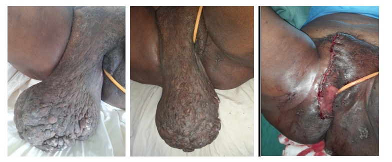
Case Report: A 70-year post-menopausal woman from Asmara presented with genital swelling which grows gradually over 15 years. She had history of trans-abdominal hysterectomy for abnormal uterine bleeding before 20 years. She had also history of right sided body weakness due to cerebro-vascular accident for the last10 years. She had no known chronic diseases such as hypertension or diabetes mellitus. On physical examination, she is a wheelchair patient with right sided hemiplegia and old midline abdominal scar. On genitourinary examination, patient had a large, about 20x18cm, non-tender, irregular, nodular mass, that hangs from the right side of the labia majora to the level of one third of upper thigh. She had also multiple nodular lesions around the ano-genital area. Different laboratory baseline investigations revealed normal finding. She was operated on 30/10/2022 and a 4kg vulvar mass was resected with right partial hemi-vulvoctomy and sample was sent for pathology which revealed fibromatosis. She was discharged with full improvement and currently on follow-up for possible recurrence.
Conclusion: This is reported for its rarity and its enormously large size, slow growing and non-invasive nature. Besides, this was atypical being in a postmenopausal woman, and it’s important to highlight that desmoids tumors should be included in the differential diagnosis of any vulvar mass. She was managed with surgical intervention as it was extremely big. Enhancing community and health professionals’ awareness on similar health problems is crucial for early intervention of similar r conditions.
Pelvic fibromatosis or desmoids tumour is a rare, non-metastatic tumour that tends to be locally invasive. It is always recurrent and arises from the fascial sheath and musculoaponeurotic structures [1]. Clinically, desmoids may be mistaken for ovarian, mesenteric or retroperitoneal tumours. Desmoids have a predilection to develop in women of reproductive age [1].
Superficial angiomyofibroblastoma (AMFB) of the genital tract typically arises from the subepithelial stromal cells of the vagina, less commonly from the vulva and cervix. Clinically, these tumors are often confused with Bartholin’s gland cysts, because of their slowly growing nature and the formation of pedunculated mass on the vulva [2, 3]. The oestrogen receptor appears to be implicated in the pathogenesis of desmoid tumour, since fibromatosis is common in women of childbearing age and regresses following menopause [1].
AMFB typically measures less than 5cm; however, case reports describe tumors of up to 23 cm in size [3]. Only five cases of vulvar AMFB with pedunculated mass have been reported in the English literature and all cases involving the labia majora and middle-aged women [4]. AMFB is an important consideration in the premenopausal and perimenopausal patient presenting with a vulvovaginal mass given its predilection for this region of the female genital tract [3].
Although surgery has been the primary treatment, complete local excision is difficult and is associated with high recurrence and complication rates. To preserve the fertility of women of reproductive age, various alternative treatments have been used, including steroids and tamoxifen and toremifene [1].
To the knowledge of the researchers, there are no reported cases of fibromatosis in Eritrea.
A 70-year-old postmenopausal woman from Asmara presented with genital swelling which affect her quality of life more than a decade. The swelling increased over 15 years progressively to extend to the level of upper thigh. She had history of trans-abdominal hysterectomy for abnormal uterine bleeding before 20 years. Besides, she had history of right sided body weakness due to cerebro-vascular accident which led her to walk in wheelchair for 10 years. She had no history of vaginal bleeding, vaginal discharge, urinary complaint and urinary or stool incontinence. Besides, she had no trauma to the genital area or any change of bowel habit. She was offered physiotherapy but without much improvement. She had no history of any chronic diseases such as diabetes mellitus, hypertension or Acquired Immune Deficiency Syndrome (AIDS). Patient refused to seek medical care for her vulvar swelling, embarrassed by the vulval mass and chose to visit many traditional healers but with no successes. She had 2 weeks history of right lower leg swelling for which she was diagnosed with cellulitis and put on antibiotics. She had history of ano-genital swelling of 4 years duration.
On physical examination, she was obese patient on wheelchair, not in cardiorespiratory distress. Her vital signs revealed blood pressure of 120/70mmHg on left arm sitting position, pulse rate of 90 beats per minute, respiratory rate of 26breaths per minute, random blood sugar of 121g/dl and temperature of 36.7oc on right axilla. She had right sided body weakness and old midline abdominal scar. There was massive vulvar mass that outspreads from the right side of the labia majora to the level of the upper thigh. The swelling was about 18x20cm, non-tender with irregular surface. (Figure: 1) She had multiple nodular lesions around the ano-genital area. Right lower leg was swollen and hyperemic.
She was investigated with fasting blood sugar, complete blood count, renal and liver function test and revealed normal finding. She was non-reactive for HIV and syphilis. On gynecological examination and vaginal ultrasound, the uterus and bilateral uterine adnexa showed no abnormalities. Tumor markers of CA 125, CA-19.9, alpha fetoprotein and carcinoembryonic antigen were within the normal range. After counseling, she was operated on 30/10/2022 and a 4kg vulvar mass was resected and right partial hemi-vulvoctomy was done. Sample was sent for pathology analysis and revealed fibromatosis. Cryotherapy was also done to the ano-genital warts, and she was discharged with improvement and regular follow-up for any possible recurrence of the disease.
Microscopic Description: Section show poorly fixed specimen, featuring keratinized stratified, Epithelial unchanged by proliferative fibroblast with collusion buds. The lesion is infiltrating a fatty tissue. No necrosis and no prominent cellular atypia. Diagnosis- Fibromatosis
Pelvic fibromatosis or desmoid tumor is a rare, non-metastatic benign tumor that occurs almost exclusively in the vulvo-vaginal region of women and tends to be locally invasive. In this case it originated from the right labia majora extending to the right perineum, which was consistent with literatures [3, 5, 6, 7]. The lesion always involves the labia majora [4, 7]. The reason why it specifically involves the labia majora is not clearly defined, but it could be due to the pathogenesis of the disease and how it relates to the estrogen receptors in the premenopausal women.
This patient had the disease for 15 years and didn’t want to resort medical care in a health facility as she was embarrassed by the disease and instead visited many traditional healers. This was consistent with many literatures that most patients felt humiliated to report to the hospital early as the growth was in the genital area and associated them with sexual transmitted diseases [5]. There are reports of other studies where a painless mass has been present from a few weeks up to 13 years [4, 7]. Communities’ awareness is crucial to encourage early health seeking to decrease the recurrence of the disease and improve quality of life.
This benign tumor is an important consideration in the premenopausal and perimenopausal women. Contrary to this common notion our case was post-menopausal, 70 years of age, having had the pathology for 15 years. Literatures have reported similar findings in younger women (in 22-year-old [5], 21-year-old [7], 46-year-old [6], 30-year-old [3] and majority of cases were observed in women less than 60 years of age [3]. Other study reported that patients have an average age of 45.8 years [4, 7] and such tumors may occur at any age, though usually they are found between the ages of 20 and 50 years [8].This was consistent with the proposed pathogenesis that it is estrogen dependent. The estrogen receptor appears to be implicated in the pathogenesis of desmoid tumor, since fibromatosis is common in women of childbearing age and regresses following menopause. In our case, the age was inconsistent with the literatures and very rare to present with such massive fibromatosis in postmenopausal woman.
This fibromatosis was nodular, non-tender and slow growing, which was reliable with other literatures. They reported similar findings on the duration of the disease and clinical finding with huge, firm, nodular and non-tender genital mass with limited mobility and originating from the right labia [5, 6, 7]. This characteristic makes it difficult to differentiate from other clinical scenarios clinically without histopathology evaluation.
In this case the size of the mass was extremely large (20x18cm) with a weight of 4kg. There were other literatures reporting bigger masses (30 x 22 cm) [5], but most reported cases were smaller with the average size being 14.2 ×13.6cm [4, 6, 7]. The large size of the mass could be mainly due to the long-standing duration of the illness and could be among the largest vulvar fibromatosis reported in literature.
Even though pelvic fibromatosis is very rare, it has similar clinical presentation to different diseases as bartholins gland cyst, cystocele, inguinal hernia, leiomyoma, rectocele and cervical mass. Therefore, it should be part of the differential diagnosis in the workup of any pedunculated vulvar mass even in young women with a lesion involving the labia minora. Literatures report similar suggestion [7].
As this vulvar fibromatosis was large, surgery was done with excision. This was steady with literatures [5]. Surgery has been the mainstay of treatment for fibromatosis and in cases of recurrent tumors or widely involved tumors, radiation or chemotherapy may be attempted [1, 7]. There are also reports of Successful treatment of recurrent pelvic desmoid tumor with tamoxifen. [1] If complete resection of the mass is not done, recurrence rate is high and needs regular follow-up.
The pathology report revealed fibromatosis. This was unswerving with the clinical presentation and duration of the disease. The histologic characteristics are vital for the diagnosis of the disease, as in this case with epithelia unchanged by proliferative fibroblast with collusion buds and is infiltrating a fatty tissue with no necrosis and no prominent cellular atypia. This was similar with literature for desmoid tumor [4, 7].
This is a rare type of benign tumor which originated from the labia majora, slow growing and non-invasive. The size was extremely large with long duration of disease and was atypical being in a postmenopausal woman, and desmoid tumors should be included in differential diagnosis of any vulvar mass. Surgical excision was done, and the pathologic result was reliable with the clinical course of the disease. Clinicians should be aware of this rare disease which can be associated with recurrence if there is incomplete excision, and it can be very embarrassing to the patient affecting her psychosocial well-being and overall health.
Acknowledgments: Authors acknowledge the patient for using her data
Funding: Not applicable
Competing of interest: Authors didn’t have any conflict of interest to disclose
Ethical approval: Written informed consent was obtained from the patient to present in case report
Author’s contribution: All authors have contributed on data analysis, interpretation and writing