
Research Article | DOI: https://doi.org/10.58489/2836-6204/002
*Corresponding Author: Isaac John Umaru
Citation: I. J. Umaru, Yakubu. O. E, Maryam. U. A, I. Ezekiel, Kerenhappuch. I. U, Emochone. R. Y, Silas Verwiyeh. T, V. Ifeoluwa. A, C. S. Ezeonu, Ebenezer M. A, S. O. Asuelimen, (2022). Evaluation of Antifungal Potential and Gastroprotective Effect of Ethanol Seed Extract of Spathodea Campanulata of Indomethacin Induced Gastric Ulcer, A Possible Application for Veterinary Animal. Research of Gastric Management and Hepatology. 1(2). DOI:10.58489/2836-6204/002
Copyright: © 2022 Isaac John Umaru, this is an open access article distributed under the Creative Commons Attribution License, which permits unrestricted use, distribution, and reproduction in any medium, provided the original work is properly cited.
Received: 09 September 2022 | Accepted: 20 September 2022 | Published: 04 November 2022
Keywords: Evaluation, Antifungal Gastroprotective Ethanol, Seed-Extract, Spathodea Campanulata, Indomethacin, Gastric Ulcer, Veterinary, Animal
Abstract
The present study evaluates the effects of the plant seed extract of Spathodea Campanulata administration on microbial (Aspergillus niger, Aspergillus flavin, Candida tropicalis and Fusarium oxysporium.) and ulcer inhibition on common Veterinary Albino rats.Spathodea campanulata is an African plant which is used in tropical and subtropical areas for ornamental purposes as well as for the treatment of a range of diseases such as wounds, skin rashes, and haemorrhoids.
Materials and Methods: Indomethacin for gastric-ulcer induction, Omeprazole and Tetracycline as standard drug, Disc diffusion method for antifungal and Histopathology for ulcer index.
Results: The effects of ethanol seeds extract were studied for antifungal and antiulcer. Where a significant activity observed in various concentration on different fungi indicating a positive agent for the selected pathogens. Also, with 40 albino rats over a period of 7 days, the rats were divided into five groups, those in group one served as the control, group two as the negative control (Indomethacin), group three as the positive control (Omeprazole) and group four and five are dosed ranged groups from 200-400mg/kg extract. Tissue injury and its infiltration were examined and scored; the length and breadth of the lesion were measured for ulcer index. Inflammatory and fibrosis alteration were present in ulcer treated rats, at days 7 colonic wall thickening and fibrotic remodelling were evident as well as blood vessels remodelling. The percentage rate of healing was 10.03% at 200 ppm and 25.51% at 500ppm. The present findings provide an integrated view of ulcer healing processes occurring in the ulcer induced colonic compartment of rats induced by indomethacin.
Conclusion: These ulcerogenic alterations may represent a suitable basis for understanding the effects of Spathodea campanulata ethanol seeds extract to ulcer treatment as well as an antifungal.
One of the major problems in veterinary animals around the world is infectious disease, such as diseases brought on by pathogens. In particular, ulcers, gastrointestinal disorders, and fungal illnesses are prevalent in cows, sheep, rabbits, and goats all the time.[1] Therapies using these generic medications resulted in a number of problems, including the infections' increased resistance to treatment and the ulcers' inability to heal. This seems to be more common in canines, equines, sheep, rabbits, and rats.[2] The African plant Spathodea campanulata is utilized in tropical and subtropical regions for ornamentation and the treatment of a number of ailments.[3] In many nations, including Ghana, Nigeria, Uganda, Gabon, Cameroon, and Senegal, Spathodea campanulata is a common plant. In Ghana, a decoction made from the Spathodea campanulata leaves and stem bark is traditionally used to treat various ailments, such as skin rashes, hemorrhoids, and stomach ulcers.[4] Both where it is native and where it has been imported, Spathodea campanulata has several medical benefits. Bark, leaf, and flower extracts are used as antidotes for poisons as well as to treat oedema, diarrhea, constipation, gastrointestinal disorders, ulcers, skin illnesses, wounds, fever, and urethral inflammation. As a preventative for malaria, it might be useful. [5-6]
The use of plant and plant extracts in rabbit husbandry appears to give a reasonable means of enhancing the welfare and health of veterinary animals, according to prior in vitro studies. Accordingly, the current study sought to assess the antifungal capability and gastroprotective impact of an ethanol seed extract of the plant Spathodea campanulata on indomethacin-induced stomach ulcers as a prospective application for veterinary animals.
Refrigerator, mortar and pestle, Whatmann filter paper no. 4, beakers, hand glove, weighing scales Paraffin wax, buffered formalin, Haematoxylin and eosin, chloroform anesthesia, ethanol, orogastric cannula, and microscope
Plant material
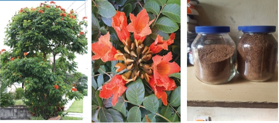
Fresh roots of Spathodea campanulata were collected from the federal University wukari Taraba State, Nigeria. The plant Spathodea campanulata seed as shown in figure 1 (African tulip tree) was dried under room temperature in open space in Biochemistry laboratory.
Preparation of plant material
The seed of the plant Spathodea campanulata were washed with distilled water to remove dust particles they were thoroughly air dried and powdered using pestle and mortar and extraction was done using ethanol by placing 150g of the powdered samples into an Erlenmeyer flask and ethanol three times the weight of the extracts was added, the solution was covered and shaken at an interval of an hour and then allowed at room temperature to stand for 7days, the mixture were then filtered using whatman filter paper No.4 and the solvent was evaporated using a rotary evaporator under reduced pressure below 50EC. It was then stored in a refrigerator until required.
Breeding of animals (albino rats)
Forty male albino rats weighing between (150-190g) were obtained from the animal farm, National Research Institute Vom, Jos Plateau State Nigeria. They were put in cages at room temperature (20-27oc) under 12/12 night/dark. They were maintained on a standard animal pellet (vital feeds, Grands cereals and oil meal Jos) and water ad libitum.
Chemical and reference drug
All chemicals used in this investigation were of analytical grade and were obtained from SIGMA Omeprazole (reference drug) was obtained from a pharmacy shop in Jos plateau State, Nigeria. The drug is an anti-ulcer drug which blocks the enzymes in the wall of the stomach from producing acid, the main culprit in peptic ulcer. By blocking the enzymes, the production of stomach acid is decreased, thus allowing the ulcer to heal.
Experimental design
The albino rats were randomly divided into five groups, this include normal group, negative and positive control group while two groups for extracts dosage, except the normal all the groups were induced with ulcer, the animals were starved from food 24hours and water 2hours before the commencement of the experiment.
Group 1: Normal control (diet/water)
Group 2: Rats (induced ulcer indomethacin 25mg/kg/bwt +diet /water)
Group 3: Rats (induced ulcer indomethacin 25mg/kg/bwt +diet/water + Omeprazole)
Group 4: Rats (induced ulcer indomethacin 25mg/kg/bwt +diet /water+100mg/kg/bwt extracts)
Group 5: Rats (induced ulcer indomethacin 25mg/kg/bwt +diet /water+/kg200mg /bwt extracts)
Group 6: Rats (induced ulcer indomethacin 25mg/kg/bwt +diet /water+300mg/kg/bwt extracts)
Group 7: Rats (induced ulcer indomethacin 25mg/kg/bwt +diet /water+400mg/kg/bwt extracts)
Group 8: Rats (induced ulcer indomethacin 25mg/kg/bwt +diet /water +500mg/kg/bwt extracts)
Experimental procedure
Animals were divided into eight groups of 5 rats each that had fasted for 24 h prior to orally treat with 25mg/kg/bwt of indomethacin for gastric-ulcer induction. Indomethacin was administered intragastrical via the aid of an orogastric canula. After 50min, an oral dose of saline, (Omeprazole, 20 mg/kg) and ethanolic seed extract of Spathodea campanulata (100, 200 300, 400, and 500 mg/kg). After 1 hour of administration of ethanolic seed extract of Spathodea campanulata, the animals were sacrificed by chloroform anesthesia. The animals were dissected and the stomach was taken out. Finally, the ulcers were observed microscopically. The observation was made for any bulging or inflammation in the stomach. By sacrificing the rats, stomach was removed and opened along the greater curvature and washed it slowly under tap water, but it on the glass slide and observed with naked eye. The stomach was carefully keeping the esophagus closed, opened along the greater curvature and the gastric contents were removed. The mucosa was flushed with saline and observed for gastric lesions using the microscopic structure.
Determination of ulcers lesions
The extents of lesions were measured in form of Ulcer Area (UA). The lengths of ulcer on the gastric mucosa were measured using a plane square ruler. The Ulcer Area (UA) was calculated. The percentage (%) of protection (P %) availed to the animals through the various treatments were calculated using the formula:
P% = (UA ulcer control - UA treatment) × 100/UA ulcer control
Histopathology
The stomach Histopathology of all the animals were fixed in 10% buffered formalin in labeled bottles and processed routinely for histology examination. The tissue embedded in paraffin wax were sectioned in 5µm thick, stained with Hematoxylin and eosin, mounted on glass slides and then examined under a standard light microscope.
Fungal culture
Authentic Clinical isolated pure culture of fungi Aspergillus niger, Aspergillus flavin, Candida tropicalis and Fusarium oxysporium were obtained from Microbiology Department, Federal University Wukari. All the strains were confirmed by cultural and biochemical characteristics and maintained in slants for further use.
Evaluation of antifungal activity of Spathodea campanula
Antifungal activity of ethanolic Seed extract of Spathodea campanulata was determined using a modified Umaru et al. [8] Disc diffusion method briefly, 100 μl of the test fungi were grown in 10 ml of fresh media until they reached a count of approximately 108 cells/ml, 100 μl of fungal suspension was spread onto the Nutrient agar plates. The extracts were tested using 5 mm sterilized filter paper discs. Discs were impregnated with (25 μg/ml, 50μg/ml, 100µg/ml, 200µg/ml, 300µ/ml and 500µg/ml) of the test samples (ethanol seeds extract), allowed to dry and placed onto inoculated plates (30 min incubation). The plates were allowed to stand at 4 0C for 2 hours before incubation with the test fungal agents. Plates inoculated with Aspergillus niger, Aspergillus flavin, Candida tropicalis and Fusarium oxysporium were incubated at 37oC for 24 hours, then the diameters of the inhibition zones were measured in millimeters. Each antifungal assay was performed in triplicate & mean values were reported. Standard antibiotic tetracycline (30 μg/ml) served as positive control for antimicrobial activity. Filter discs impregnated with 10 μl of distilled water were used as a negative control. Solvent control disc (ethanol) was also placed with the test, positive and negative control.
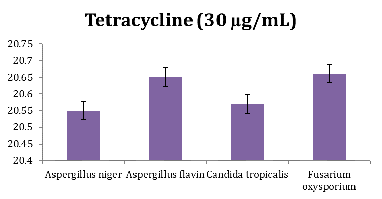
Chat 1: Antifungal activity of tetracycline (30 µg/mL) against Aspergillus niger, Aspergillus flavin, Candida tropicalis and Fusarium oxysporium
Chat 2: Antifungal activity of Ethanol extract (25 ug/ml) against Aspergillus niger, Aspergillus flavin, Candida tropicalis and Fusarium oxysporium

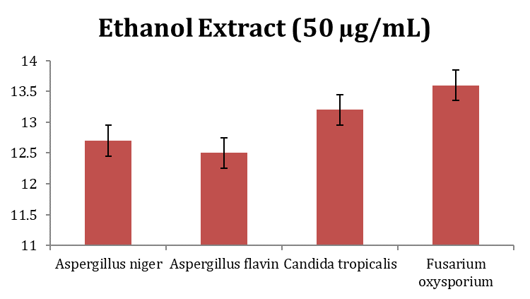
Chat 3: Antifungal activity of Ethanol extract (50 µg/mL) against Aspergillus niger, Aspergillus flavin, Candida tropicalis and Fusarium oxysporium
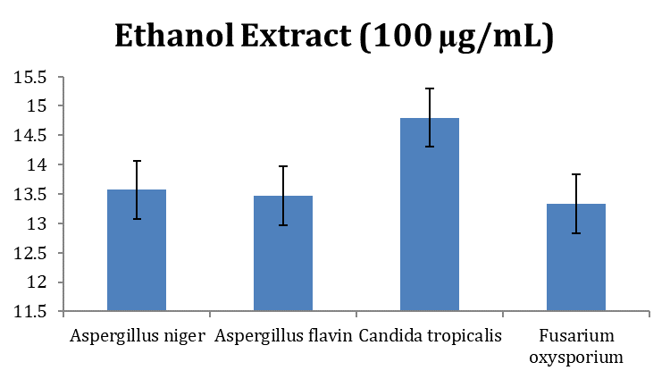
Chat 3: Antifungal activity of Ethanol extract (100 µg/mL) against Aspergillus niger, Aspergillus flavin, Candida tropicalis and Fusarium oxysporium
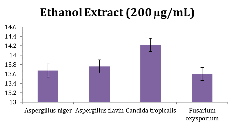
Chat 4: Antifungal activity of Ethanol extract (200 µg/mL) against Aspergillus niger, Aspergillus flavin, Candida tropicalis and Fusarium oxysporium
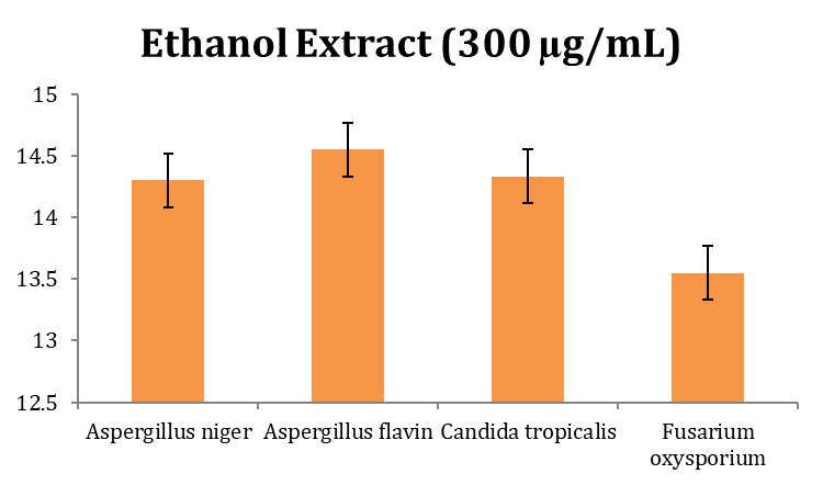
Chat 5: Antifungal activity of Ethanol extract (300 µg/mL) against Aspergillus niger, Aspergillus flavin, Candida tropicalis and Fusarium oxysporium
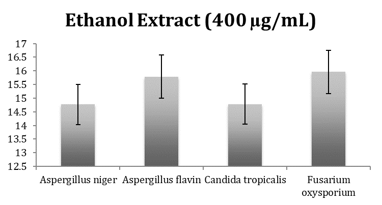
Chat 6: Antifungal activity of Ethanol extract (400 µg/mL) against Aspergillus niger, Aspergillus flavin, Candida tropicalis, and Fusarium oxysporium.
Anti-ulcerogenic effects

Figures i and ii are the normal and negative control. The figures show normal gastric and duodenal mucosal overlying muscle coat. There is focal lymphocytic inflammation and aggregate constituent with the Mucosal Associated Lymphoid Tissue (MALT) as shown in figure ii, Ulceration after induced by indomethacin Figure iii represents the effect of the ethanol extracts 7 days after treatment. At 500 mg/kg/bwt, duodenal ulcer: The section shows non-dysplastic gastric and duodenal mucosal. There is an area of ulceration at the duodenum covered by necrotic debris of inflammation. It extends up to the muscle coat.
Table 2: Effect of Ethanol Extracts from Spathodea Campanulata seeds of indomethacin induced ulcer rats and percentage protection
Group | Ulcer index | Percentage (%) |
-ve control | 39.2 | 0.00 |
+ve control | 0.12 | 86.25 |
100 mg/kg/wt | 9.34 | 8.11 |
200 mg/kg/wt | 11.12 | 10.03 |
300 mg/kg/wt | 16.44 | 12.37 |
500 mg/kg/wt | 29.20 | 25.51 |
Result is Mean + SD. N = 5
*= significant activity was observed.
Concentration of standard is 20 µg/mL of Omniprazole
This study evaluates the effect of seeds crude extract of Spathodea Campanulata in ulcer induced albino rats and non-ulcer induced albino rats. The findings of the study will help the under developed countries and the world at large in advancing the search for natural compounds that could be used either as a whole or incorporated into drugs as a remedy to stop the menace and suffering caused by ulcer in Veterinary animals.
The result shows the finding and the potentials of the ethanol extract of Spathodea Campanulata on ulcer induced Veterinary animals (Rabbits, rats, Goats, Cows etc). Table 2 shows dose-dependent inhibition of gastric ulceration at 100 mg/kg/bw, 200 mg/kg/bw, 300 mg/kg/bw, 400 mg/kg/bw, and 500 mg/kg/bw, higher inhibition value with percentage protection of 25.51% was observed when compared to Omniprozole used as the test control of 0.12 of 86.25 % protection. This activity might be due to the presence of chemical constituents in the extract. This result agrees with the report of Sabiu et al. [9] that gastroprotective potentials of plant extracts against indomethacin-ulcerated rats were associated with their various bioactive compounds present in the extract.
Recently, pathogens of various species have been incriminated in deadly cases of the perspiration epidermis in piglets and dermatitis as reported by Nemeghaire et al., [10] and Chen et al., [11] as well as mastitis in cattle. [12-14] With this in mine, the results obtained will go a long way to cutile the invasion of these pathogen among the veteneary kingdom.
Among which is Aspergillus niger is a fungus and one of the most common species of the genus Aspergillus. It causes a disease called "black mold" on certain fruits and vegetables such as grapes, apricots, onions, and peanuts, and is a common contaminant of food. It is ubiquitous in soil and is commonly reported from indoor environments, where its black colonies can be confused with those of Stachybotrys (species of which have also been called "black mold"). [15]
As described by Nemeghaire et al., [10] and Chen et al., [11], as well as dermatitis and mastitis in cattle, infections of diverse species have recently been implicated in fatal cases of the sweating epidermis in piglets and dermatitis. With this in mind, the outcomes will significantly reduce the spread of these pathogens among the veterinary kingdom. A fungus and one of the most prevalent species of the Aspergillus genus is Aspergillus niger. On several fruits and vegetables, including grapes, apricots, onions, and peanuts, it causes a disease known as "black mold" and is a frequent food contaminant. It is prevalent in soil and frequently seen in interior settings, where its black colonies can be
According to this study, Tetracycline has a higher zone of inhibition on Aspergillus flavin and Fusarium oxysporium, measuring 20.65 and 20.66 mm in Figure 2 and 25 g/mL in Figure 3. Candida tropicalis and Aspergillus niger had zones of inhibition of 14.5 mm, 14.5 mm, and 14.2 mm, respectively.
A 50 g/mL ethanol extract's antifungal activity against Aspergillus niger, Aspergillus flavin, Candida tropicalis, and Fusarium oxysporium was demonstrated in Figure 4. Aspergillus flavin has the lowest activity at 12.3 mm, whereas Fusarium oxysporium has the highest activity at 13.5 mm. With the study of Thampi and Kumar16 that the methanolic leaves extract had a considerable inhibition within 16mm and 17mm against A. niger and C. albicans with rise in concentration, we are now able to grasp the effect of this aggressive plant.
Figure 5 shows the ethanol extract's (100 g/mL) ability to prevent the growth of the chosen fungus. Candida tropicalis displayed the most activity, measuring 14.7 mm, while Fusarium oxysporium displayed the lowest activity, measuring 13.00 mm. While in Figure 6, the crude extract's activity at 200 g/mL was seen to be extremely active on Candida tropicalis only with 14.2 mm when compared to the other pathogens, confirming Thampi and Kumar's16 observation that Candida tropicalis was seen to be more inhibited at 100 g/mL with 17 mm.
When compared to Aspergillus flavin and Candida tropicalis with 20.66 and 20.67 mm, the extract at concentration 300 g/mL Figure 7 significantly inhibited Aspergillus niger, Fusarium oxysporium, and other aspergilli. The antifungal activity of ethanol seed extract (400 ug/ml) was demonstrated in Figure 8 against Aspergillus niger, Aspergillus flavin, Candida tropicalis, and Fusarium oxysporium. Aspergillus niger exhibits the lowest activity, whereas Fusarium oxysporium exhibits the highest activity, measuring 16.10mm.
The same pattern was seen in the study of Dolezal et al. (17), which described a fungus with a global distribution that Aspergillus flavin inhibited with a 15.79 mm inhibition at 500 g/ml. This fungus is well recognized for settling on tree nuts, legumes, and cereal grains. Typically, postharvest rot starts during the harvest, storage, and/or transportation. Its scientific name, flavus, refers to the yellow tint of the spores, which are frequently noticed, and comes from the Latin word for yellow. Preharvest A. flavus infections are possible, but dormancy, or lack of symptoms, frequently persists until postharvest storage and/or transport. [18,19]
The zone of inhibition for Candida tropicalis was 20.67 mm at 400 g/ml. This yeast species belongs to the genus Candida. It is a typical pathogen in neutropenic hosts, where it can disseminate to peripheral organs through the circulation. 20 Amphotericin B, echinocandins, or extended spectrum triazole antifungals are among therapies for invasive disease. [21] As a result, it was noted from the data that the zone of inhibition increases as the concentration does. [22]
At different concentrations of the crude extract of Spathodea campanulata, it was found to be active against Aspergillus niger, Aspergillus flavin, Candida tropicalis, and Fusarium oxysporium, particularly at the highest concentration of the extract at 400 g/ml against the fungi infecting veterinary animals. To treat diseases and ailments in home boundary animals for improved health and a healthier environment, this plant seed's substantial activity of the crude extract on the induced ulcer has provided a hint.
This study discovers that Aspergillus niger, Aspergillus flavin, Candida tropicalis and Fusarium oxysporium and Ulcer disease could be a major sudden death of Animal husbandry cases in Nigeria. This study will help the researcher and the veterinary personels to uncover the possible means to prevention and treatment of diseases caused by these pathogens for a better Animal domestication
We acknowledge Natural Product Research Laboratory Farm (NPRL) Federal Housing Estate Bajabure Gerie Adamawa State for the supply of this animals
The authors report that there is no conflict of interest