
Research Article | DOI: https://doi.org/10.58489/2836-5038/012
1Faculty of Medicine, Ain Shams Research Institute (MASRI), Faculty of Medicine in Shams University. Cairo, Egypt
2Zoology Department, Faculty of Science, Ain Shams University, Faculty of Medicine Ain Shams University. Cairo, Egypt.
*Corresponding Author: Fatma A. Abu Zahra*
Citation: Fatma A. Abu Zahra, (2023)., An Ultrastructural analysis of the HuH7 Hepatocellular carcinoma cell line’s response to conditioned and co-culture media. International Journal of Stem Cells and Medicine 2(1). DOI: 10.58489/2836-5038/012
Copyright: © 2023 Fatma A. Abu Zahra, this is an open-access article distributed under the Creative Commons Attribution License, which permits unrestricted use, distribution, and reproduction in any medium, provided the original work is properly
cited.
Received: 28 March 2023 | Accepted: 17 April 2023 | Published: 19 August 2023
Keywords: Hepatoma cell line (HuH7), Umbilical cord Mesenchymal Stem Cell (UMSC), proliferating cell nuclear antigen (PCNA) and Vascular Endothelial Growth Factor (VEGF).
In this study, the gene expression of some cancer-related genes was measured to differentiate between two types of therapeutic media in which hepatocellular carcinoma (HuH7) were harvested. The first was a conditional medium in which umbilical cord mesenchymal stem cells were harvested then diluted by DMEM media (1: 1) and the second was Co-culture medium that mesenchymal stem cells and HuH7 cells (1:1) together were incubated for 3 days in a medium. The latter was called Co- culture medium. Comparison of the Results of gene expression of Surviving, PCNA, Beta-Catenin, Telomerase and VEGF by using real-time PCR in several four-four-time intervals (48-72-96 hrs.) showed that in conditioned media and co-culture media, hepatoma cell line (HuH7) expressed a significant down-regulation in Surviving (p< 0.001), PCNA (p<0.001), VEGF(p<0.001) and Beta- Catenin(p=0.022) but not significant in Telomerase (p= 0.617). In our study, the measure of gene expression at different time (ALT), Aspartate aminotransferase (AST), Albumin, Alkaline phosphate, gamma GT, alpha- fetoprotein when compared regarding time Results in a significant in Alpha-fetoprotein (p < 0.001), ALT (p < 0.001) and AST (p < 0.001) while the results of Albumin (p = 0.650), Alkaline phosphatase (p = 0.329) and Gamma GT (p = 0.844) were non-significant.
Hepatocellular carcinoma (HCC) represents the fifth most common malignant tumor and the third most common cause of mortality related to cancer in the world (1). HCC is cancer that has a high number of risk factors, such as viral infection by either virus B or C virus, alcohol, or prolonged aflatoxin exposure (2). Umbilical cord mesenchymal stem cells retain their intrinsic stem cell potential while at the same time displaying high proliferation rates, low immunogenicity, and powerful capacity to differentiate into a wide range of lineages including osteocytes, adipocytes, chondrocytes, and neural lineages (3). So, UC-MSCs represent excellent candidates for cell therapy in regenerative medicine and tissue engineering. Many preclinical researches on different disease models have shown that-MSCs can accelerate wound recovery and suppress tumor progression. UC- MSCs possess a higher therapeutic potential than MSCs derived from other tissues.
Increasing evidence suggests that the mechanism underlying UC-SMCs efficacy depends mostly on cell secretions, in contrast to the early paradigm of codifferentiation capacity. They exhibit replacement and differentiation (4). It has been shown that MSCs can produce a vast array of cytokines and extracellular molecules and express receptors and/or counter receptors both for cell–cell, and cell–matrix interactions (5 and 6). More recently, MSCs have become a topic of great focus in relation to cancer. Growing evidence suggests that MSCs home to not just injured tissues but tumors (7), where they have an important role in affecting the behavior of tumor cells. Thus, the ease of MSCs isolation and the fact that they can be home to wound or cancer sites make them a potential platform for cell-based targeting delivery. In response to liver injury or loss of liver mass, the proliferation of mature liver cells is the first-line defense to restore liver homeostasis (8). Mesenchymal stem cells (MSCs) play several simultaneous roles to limit inflammation through releasing cytokines, and aiding healing by expression of growth factors (8). The aim of this study was to investigate the effect of hMSCs derived from the umbilical cord on the growth of HepG2 to explore the anti-proliferative and the tumor suppressive effect of conditioning and co-culturing umbilical cord mesenchymal stem cells with hepatocellular carcinoma on human hepatoma cell line (HepG-2). The study also aimed to investigate the regressive changes in the ultrastructure of HepG2 cells using transmission electron microscopy.
Cell and tissue material preparation: This work was done in the Faculty of Medicine Ain Shams Research Institute (MASRI), Tissue Culture Unit. Mesenchymal Stem Cells from Human Umbilical cords Wharton’s Jelly (n = 12; gestational ages, 39-40 weeks) were collected and processed within 2-3 hours after Caesarean births attending the Women's Hospital and Obstruction of Faculty of Medicine Ain Shams University.
Isolation of Mesenchymal stem cell from Umbilical Cord (UC)
UCs were washed with Dulbecco’s PBS several times, cut into small pieces, and filled with 0.1% collagenase type П for cell digestion (Sigma-Aldrich, St. Louis) in PBS and shaken in the water bath at 37°C for 60 min. Each UC was washed with proliferation medium, and the detached cells were harvested after gentle massage of the UC. Cells were centrifuged at 300 xg for 10 min, re-suspended in proliferation medium, and seeded in 25- cm3 flasks (Nunclon, Germany) at a density of 5×10⁵ cells per ml in complete media. After 72 hrs..... of incubation in a humidified atmosphere with 5% CO2, non-adherent cells were removed, and the culture medium was replaced every 3 days. On day 14, the adherent colonies of cells were almost confluent and removed by using trypsin EDTA, and counted. Cells were identified as being MSCs by their morphology, adherence, and their power to differentiate into osteocytes, neurocytes, and chondrocytes.
Flow cytometry identification of UC- MSC surface biomarkers.
UC-MSCs were used for cell identification, cells were washed with PBS and digested with 0.25% trypsin. After centrifuging at 300 x g for 5 min, the supernatant was removed and cells were washed twice with PBS. Cells were then incubated with FITC-conjugated primary antibodies against CD19, CD44 and CD105 for 30 min at room temperature in the dark. The expression of cell surface biomarkers was determined using flow cytometry analysis. Fluorescence acquisition was performed in a FACS Calibur II cytometer (Becton Dickinson, San Jose, CA, USA) (9).
Cell Culture of human hepatoma (HuH7) cell line:
Human Hepatoma (HuH7) cells in Egypt (Japanese male type collection) and grown in a sterile 75-cm2 tissue culture flask in a complete medium containing DMEM supplemented with 10
RNA extraction and real-time PCR
Total RNA from treated and untreated HuH7 cells were harvested and washed with PBS at 4°C. Total RNA from cell cultures was extracted using a total RNA extraction Kit supplied by Qiagen, according to the manufacturer’s instructions. All RNA samples were treated with DNase I at 37°C for 30 min for removal of any genomic DNA contamination. cDNA was generated from 500 ng of total RNA extracted with 1 μl (20 pmol) antisense primer and 0.8 μl superscript AMV reverse transcriptase at 25°C for 10 min, 37°C for 120 min, and 85°C for 5min and finally 4°C to infinity. The relative abundance of mRNA species was assessed using the SYBR® Green method on an ABI prism 5700 fast (Applied Biosystems, Foster City, CA, USA). The reverse transcription (RT) product was used to measure the expression of glyceraldehyde 3-phosphate dehydrogenase (GAPDH) as a positive housekeeping gene, the mRNA
expression of PCNA, Survivin, β –catenin, VEGF, and Telomerase was done using real-time PCR (qPCR), and PCR primers were designed with Gene Runner Software (Hasting Software, Inc., Hasting, NY) for RNA sequences from GenBank (Table 1).
Table 1: Primers sequences used in this study (F, forward; R, reverse)
| Gene | Primers |
PCNA (GenBank Accession: G08890) | Forward: 5'-AAA AAA GAC TAT GAA GTG GGT AGG-3' Reverse: 5'-CTG TTT CTA CAG TGC ATT GTA TAC G-3' |
Survivin (GenBank Accession: NM 001168) | Forward: 5'-ATG GCA CGG CGC ACT TT-3' Reverse 5'-TCC ACT GCC CCA CTG AGA A-3' |
β –catenin (UniGene Hs:166011) | Forward: 5'-GGA AGG TGG GAT TTT TGG TT-3' Reverse 5'-TCC TCT TCT GCT CTT TTC TTG G-3' |
VEGF (UniGene Hs:73793) | Forward: 5'-CTG CTG TCT TGG GTG CAT TG-3' Reverse: 5'-TTC ACA TTT GTT GTG CTG TAG-3' |
Telomerase (UniGene Hs: 492203) | Forward: 5'-ACT CGA CAC CGT GTC ACC TA-3' Reverse: 5'-GTG ACA GGG CTG CTG GTG TC-3' |
GAPDH (UniSTS: 273246) | Forward:5'-GAAGGTGAAGGTCGGAGTCA-3 Reverse:5'- GAAGATGGTGATGGGATTTC-3' |
All primer sets had a calculated annealing temperature of 60°. Quantitative RT-PCR was performed in duplicate in a 20 µL reaction volume consisting of 2X SYBR Green PCR Master Mix (Applied Biosystems), 900nM of each primer, and 2-3µL of cDNA. Amplification conditions denaturation at 95 °C for 15 min, followed by 45 cycles each cycle at 94°C for 15 sec, 60°Cfor 25 sec, and 72°C for 20 sec. Data from real-time assays were calculated using the v1·7 Sequence Detection Software from PE Biosystems.
(Foster City, CA, USA). Relative expression of Surviving PCNA, β-Catenin, Telomerase, and VEGF mRNA was calculated using the comparative Ct method as previously described. All values were normalized to the glyceraldehyde-3-phosphate dehydrogenase (GAPDH) gene 2-∆∆Ct method [∆Ct= Ct (gene) - Ct (GAPDH)] and reported as fold change (ΔΔCT) over background levels detected. Melting curves were performed to ensure that only a single product was amplified.
Statistical analysis
The statistical analysis in this study was performed using the following programs: IBM SPSS 22.0.0.2. Python packages (Pandas 0.20.3 and SciPy 0.19.
In the present study, the hepatoma cell line HuH7 was chosen as a model for liver cancer. The main objective of the study was to evaluate the effect of two different media, conditioned media (the medium in which umbilical cord mesenchymal cells were cultured) and co-culture medium (the medium in which mesenchymal and HuH7 cells were cultured) on different parameters in HuH7 cultured cells. Mesenchymal stem cells from cord (Wharton’s jelly) were also investigated at 0,3,10 and 14 days from culturing where the cells reached 95-100% confluence after 14 days (Fig. 1).
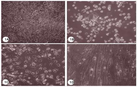
Fig. 1: Photomicrographs showing growth of mesenchymal stem cells from the cord (Wharton’s jelly) (Inverted microscope, X400).
In 0 time from culture, B) 3 days after changing media, C) 10 days from culture, D) 14 days showing 95-100% confluence of cells.
Culture of HuH7 cancer cells with hMSC-conditioned media
To determine whether the cultured hMSCs release soluble factors that can affect cancer cell growth, we cultured HuH7 in hMSCs-conditioned media. After 48hrs., the number of dead cells was increased and there were changes in shape that’s because conditioned media derived from hMSCs could inhibit cell growth relative to control cells (Fig. 2).

Fig. 2: Photomicrographs showing the effect of conditional media on hepatocellular carcinoma (HuH7) (Inverted microscope, X400). A-Control (confluent HuH7), B- 48 hours after treatment, C- 72 hours after treatment, and D-96 hours after treatment.
The results were generally in agreement with those obtained by the Trypan Blue assay, in which the conditioned media exerted an inhibitory effect on cell growth. After 72 hrs., the number of dead cells increased and cells were damaged while, after 96hrs. the larger number of dead cells was observed. After treatment of HuH7 cells with co-culture media (Fig. 3), for 48hrs. a number of dead cells appeared and cells were damaged, after 72 hrs. the number of dead cells increased and cells were more dissociated while, after 96hrs. Most of the cells appeared dead and severely damaged.
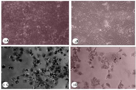
Fig. 3: Photomicrographs showing effect of co-culture media on hepatocellular carcinoma (HuH7) (Inverted microscope, X400).
(A) Control (confluent HuH7), (B) 48 hours after treatment.
(C) 72 hours after treatment, (D) 96 hours after treatment.
Cell Viability assay (MTT)
By using the MTT assay, it was found that there were highly significant differences in the viability between the hepatoma cell line (HuH7) cultured in normal media, the hepatoma cell line (HuH7) cultured in the conditioned media (p0.001). By comparing the cell viability at the four-four-four-time intervals regarding the non–normally distributed variables, there was a highly significant difference between the four groups (0 time (HuH7 as control), 48,72 and 96hrs.). The data in the two box plots (Fig. 4) showed that the cell viability was inversely proportional with increased
| Test | Media | F | pvalue |
| Alfa- fetoprotein | Conditioned | 8.359 | 0.001 |
| Co-Culture | 6.914 | 0.01 | |
| Albumin | Conditioned | 4.401 | 0.009 |
| Co-Culture | 0.591 | 0.650 | |
| GOT | Conditioned | 29.012 | 0.001 |
| Co-Culture | 23.370 | 0.001 |
time after 96hrs. The cells showed the lowest value.
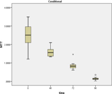

Fig. 4: MTT box plot with time for conditioned media and Co-culture media.
To compare the metabolic status of the HuH7 cells treated with UC-MSCs conditioned medium or the co-culture system, the albumin production, as well as the activity of the liver-specific enzymes AST, ALT, and GGT, were measured. There was a non-significant difference in albumin between the two groups (conditioned and co-culture groups) (p=0.054) but, in the conditioned media, comparing the four groups regarding the normally distributed variables, there was a significant difference in albumin between the four groups (0 times, 48, 72 and 96 hrs.) with a (p = 0.009). On the other hand, in the case of co-culture media, there was a non-significant difference (p = 0.650) between the four groups.
Regarding αFP levels in HuH7 cultured in normal medium and those cultured in conditioned and co-culture media, there was an obvious change. There was a highly significant difference between groups at the different four-four-time intervals. By comparing αFP in cells cultured in normal media and those cultured in conditioned and co-culture media it was inversely proportional with decrease in αFP by increasing the time of incubation.
Table 1: showing normal variable data relation with time in conditioned and co-culture media
| Test | Media | F | pvalue |
| Alfa- fetoprotein | Conditioned | 8.359 | 0.001 |
| Co-Culture | 6.914 | 0.01 | |
| Albumin | Conditioned | 4.401 | 0.009 |
| Co-Culture | 0.591 | 0.650 | |
| GOT | Conditioned | 29.012 | 0.001 |
| Co-Culture | 23.370 | 0.001 |
As for liver enzymes (GOT & GPT) there was a highly significant difference between the four-four-time intervals regarding GOT activity in the case of groups cultured in conditioned media (p<0>p0.001). Unfortunately, there was no significant difference in GPT activity in corresponding of the four-time interval groups (0-time, 48, 72, and 96 hrs.) in conditioned media (p <0>p = 0.650) between the four groups. Regarding αFP levels in HuH7 cultured in normal medium and those cultured in conditioned and co-culture media, there was an obvious change. There was a highly significant difference between groups at the different four-four-time intervals. By comparing αFP in cells cultured in normal media and those cultured in conditioned and co-culture media it was inversely proportional with a decrease in αFP by increasing the time of incubation.
Table 2: Showing comparison between the four groups regarding the non-normally distributed variables in two media
| Test | Media | X2 | p-value |
| GPT | Conditioned | 0.126 | 0.647 |
| Co-Culture | 0.112 | 0.674 | |
| Alkaline Phosphatase | Conditioned | 3.712 | 0.294 |
| Co-Culture | 5.706 | 0.125 |
Gene expression of cancer related genes.
Survivin: by comparing the different time interval in both groups, there was highly significant difference between them (0- time (HuH7 as control), 48, 72 and 96 hrs.) in Survivn RQ (ΔΔCT) for Conditioned media (p<0>
PCNA: There was a high significant difference between the two groups (conditioned and co-culture groups) in PCNA (p=0.001). Comparing the four intervals of time. groups, there was significant difference between the four groups (0, 48, 72 and 96 hrs.) in PCNA for Conditioned media (p=0.001).
β-Catenin: By Comparing the two groups regarding the nonnormally distributed variables, there was significant difference between the two groups (conditioned and co-culture groups) in β-Catenin C (p<0>
Telomerase: By Comparing the two groups regarding the non-normally distributed variables, there was a nonsignificant difference between the two groups (conditioned and co-culture groups) in telomerase C (p=0.617 and p=0.611respectively). Comparing the four groups regarding the non-normally distributed variables, there was a significant difference between the four groups (0, 48, 72, and 96 hrs.) in telomerase for Conditioned media (p <0>
Table 4: Showing comparison between the four-four-four-time intervals in the two types of media.
| Test | Media | X2 | p-value |
| Survivin | Conditioned | 40.973 | 0.001** |
| Co-Culture | 40.986 | 0.001** | |
| PCNA | Conditioned | 40.977 | 0.001** |
| Co-Culture | 40.846 | 0.001** | |
| β-Catenin | Conditioned | 40.969 | 0.001** |
| Co-Culture | 40.983 | 0.001** | |
| Telomerase | Conditioned | 39.974 | 0.001** |
| Co-Culture | 40.980 | 0.001** | |
| VEGA | Conditioned | 41.015 | 0.001** |
| Co-Culture | 36.921 | 0.001** |
VEGF
: By Comparing the two groups regarding the non-normally distributed variables, there was a significant difference between the two groups (conditioned and co-culture groups) VEGF (p<0>
MSCs have received increased attention for their ability to interact with cancer cells and make an impact on cancer cells’ behavior. However, their potential to exhibit promotional or inhibitive properties by providing the cancer cells with a microenvironment remains controversial. Some reports suggested that they exhibit a potent promotional effect by providing growth signals, while others suggested they have potential role in inhibition of the proliferation of cancer cells. The exact mechanism of their action in the regulation of cancer cells has not been well elucidated yet.
In the present study, the effect of conditioned media and co-culture media on cultured HuH7 cells was studied by measuring; morphological changes, cell viability, certain biochemical changes, expression of cancer- related genes. The results of this study showed morphological changes and dramatic decrease in cell viability which were all time dependent on the number of dead HuH7 cells which were cultured in conditioned media and co- culture media. El-nahrawy et al., showed morphological changes and possible beneficial effect of human umbilical cord MSCs- conditioned media on liver malignant cells HepG2 via the secretion of soluble factors or cytokines like IL-10 having anti-inflammatory and possible anti-tumor effects (11). Ramasamy et al., studied the effect of MSC on the proliferative activity of malignant cells of different lineages. The tumor cell lines of hematopoietic (BV173, K562, Jurkat, KG1a and wS9-B-LCL) and non-hematopoietic (UCH10 and CC3) origin were cultivated, at different ratios, in the presence of MSC and tested for their proliferative activity after 3 days of co-culture. MSC exhibited a dose-dependent antiproliferative effect on all cell lines investigated (12).
Alpha-fetoprotein (αFP) is a good tumor marker that is elevated in 60–70% of patients with hepatocellular carcinoma. Normally, levels of αFP are below 10 ng/ml, but marginal elevations (10–100) are common in patients with chronic hepatitis. The progressive elevation of alpha-fetoprotein ≥7 ng/mL/month in patients with liver cirrhosis is useful for the diagnosis of hepatocellular carcinoma in patients that do not reach αFP levels ≥200 ng/mL (13). Also, the serum concentration of αFP >400 ng/ml is considered a diagnostic marker for HCC. Although, less than half of HCC patients may generate those high levels of αFP (14). On the contrary, some investigations showed that αFP cannot be used in the initial screening of HCC due to its low sensitivity at diagnostic values. In the present study αFP values were inversely proportional with the increase of time intervals of incubation of HuH7 cells whether in conditioned media or co-culture media as they showed highly significant decrease with the lowest values recorded after 96hrs.
In clinical practices, liver functions have long been considered to assume crucial status in the prognosis of many types of cancers such as gallbladder and colorectal cancers. Liver functions can be reflected by markers not only like ALB, GELO, and TP, indicators of the nutritional status, but also ALP, ALT, AST, γ–GT, LDH, TBIL, and DBIL, indices reflecting liver damage. All these liver function parameters were firstly evaluated for their effect on the overall survival in intrahepatic cholangiocarcinoma (ICC) patients. Therefore, these liver enzymes may serve as valuable predictive markers in HCC patients (15). To date; studies have shown those abnormal changes of liver enzymes often lead to poor prognosis in a multitude of cancers. Likewise, other liver function markers such as ALP and gamma glutamyl transpeptidase (γ-GT) were often Alpha-fetoprotein (αFP) is a good tumor marker that is elevated in 60–70% of patients with hepatocellular carcinoma. Normally, levels of αFP are below 10 ng/ml, but marginal elevations (10–100) are common in patients with chronic hepatitis. The progressive elevation of alpha-fetoprotein ≥7 ng/mL/month in patients with liver cirrhosis is useful for the diagnosis of hepatocellular carcinoma in patients that do not reach αFP levels ≥200 ng/mL (13). Also, the serum concentration of αFP >400 ng/ml is considered a diagnostic marker for HCC. Although, less than half of HCC patients may generate those high levels of αFP (14). On the contrary, some investigations showed that αFP cannot be used in the initial screening of HCC due to its low sensitivity at diagnostic values.
In the present study αFP values were inversely proportional with the increase of time intervals of incubation of HuH7 cells whether in conditioned media or co-culture media as they showed highly significant decrease with the lowest values recorded after 96hrs. In clinical practices, liver functions have long been considered to assume crucial status in the prognosis of many types of cancers such as gallbladder and colorectal cancers. Liver functions can be reflected by markers not only like ALB, GELO, and TP, indicators of the nutritional status, but also ALP, ALT, AST, γ–GT, LDH, TBIL, and DBIL, indices reflecting liver damage. All these liver function parameters were first evaluated for their effect on the overall survival in intrahepatic cholangiocarcinoma (ICC) patients. Therefore, these liver enzymes may serve as valuable predictive markers in HCC patients (15). To date; studies have shown those abnormal changes of liver enzymes often lead to poor prognosis in a multitude of cancers. Likewise, other liver function markers such as ALP and gamma-glutamyl transpeptidase (γ-GT) were often risk factors in patients with some types of cancers (16).
In the present study it was found that ALP did not show any significant change in case of using co-culture media or conditioned media from 0 time until 96hrs. In tissue, ALP is well known as a membrane-bound ectoenzyme, used as an indicator to reflect hepatobiliary or bone diseases and that it is attached to the membrane via glycan phosphatidylinositol (GPI) anchor (17).
The data of supported the clinical observations recorded in numerous clinical studies that described the close relationship between the worsening of hepatocellular carcinoma with increased GT levels. The data of the present study showed highly significant decrease in the level of γGT with the increase of incubation time from 0 – 96 hrs. in case of using conditional media or co- culture media for treatment of HuH7 cells, which means according to the report of that these media have antitumor effect (18). The present data also showed lower levels of GOT and GPT after the incubation in conditional media or co- culture media at 48, 72, 96 hrs. This confirms the conception that conditioned media and co-culture media have antitumor effect on HuH7 cell line. Angiogenesis is a complex multiple- step process, which involves endothelial cell proliferation, migration, tube formation, vascular network reorganization and stabilization (19). Our findings demonstrate here that conditioned and co-culture medium of hMSCs has a suppressive effect on cell growth, depending on time-response. This distinctive property of hMSCs is probably related to the competition of production of a combination of cytokines with various effects in culture. Data from this study might provide some clues for the antitumor activity of conditioned and co-culture medium of hMSCs can avoid untoward effects of administered MSCs. Further functional investigation of the cellular and molecular events will enable the improved understanding of the mechanism of the action between cancer cells and hMSCs for development of effective therapy in the treatment of cancers. Analysis of proportion of cells in conditioned and co-culture medium of hMSCs showed that HepG2 cells were decreased in the percentage of cell population by time. The screen for the levels of cell expressing population in the mixture showed that decreased percentage of cancer cell population was apparently in presence of hMSCs. This clearly indicated that cell proliferation could be regulated by hMSCs as a result of interaction between hMSCs and cancer cells. Consistent with these results, as revealed by RT-PCR analysis, hMSCs treatment caused a marked higher or lower level of key molecules, such as Survivin, β –catenin, PCNA, Telomerase and VEGF.
Survivin is an inhibitor of apoptosis protein, which is highly expressed in most cancers and associated with chemotherapy resistance, increased tumor recurrence, and shorter patient survival, making antisurvivin therapy an attractive cancer treatment strategy. Increased survivin expression was observed in the vast majority of cancers. These included esophageal, lung, ovarian, central nervous system, breast, colorectal, bladder, gastric, prostate, pancreatic laryngeal, uterine, hepatocellular, and renal cancers, as well as melanoma and soft tissue sarcomas (20). The current study suggests that UCUC-MSCs induces apoptosis of HuH7 cells, which is associated with downregulation of survivin mRNA expression. Survivin is a member of the inhibitor apoptosis protein (IAP) family. These are bifunctional proteins that suppresses apoptosis and regulates cell division (21). The expression of survivin is highly cancer-specific and is one of the top four transcripts uniformly upregulated in human cancers, but not in normal tissues (22). The overexpression of survivin appears to correlate with aggressive tumor behavior and poor prognosis in hepatocellular carcinoma (23). So, survivin has become an ideal target for the diagnosis and treatment of cancer. Other studies have shown that overexpression of survivin was closely related to chemo resistance, and inhibition of survivin improved the sensitivity of tumor to chemotherapy (24).
Survivin is essential for cell-cycle progression in hepatocellular carcinoma cells, and down-regulation of survivin expression may lead to programmed cell death (25), indicating that survivin may be an appealing new target for novel therapies in hepatocellular carcinoma (26). Another explanation was introduced by Dai et al., who indicated that the anti-apoptotic survivin molecule may promote S-phase entry, and cooperate with the G2/M checkpoint to regulate proper cell cycle progression? (27). In the present study survivin gene is expressed at a very high rate at the beginning of experiment (0-time) when HuH7 cells were incubated in conditioned media and co-culture media survivin decreased significantly with the increase of time to 96 hrs. where it showed the lowest expression. Our results about survivin expression are also in agreement with Yang et al., who reported that UCUC- MSCs significantly inhibit the downregulating anti-apoptotic proteins Bcl-2 and surviving (28).
Cell growth, the cell cycle and apoptosis are closely associated with the genesis and development of liver cancer, and multiple factors are involved in their regulation, including surviving, caspase-3 and proliferating cell nuclear antigen (PCNA) which is associated with vital cellular processes (29 and 30).
The proliferating cell nuclear antigen (PCNA) is a nuclear protein which was independently discovered by Miyachi et al., as PCNA (31). PCNA is the heart of many essential cellular processes such as DNA replication, repair of DNA damage, chromatin structure maintenance, chromosome segregation and cell-cycle progression and can be regarded as one of their common integrators (32).
Recent studies have reported that tumor cells express increased levels of PCNA, identifying it as a potential target for cancer therapy (33). The increased expression of PCNA could have a predictive and prognostic value. However, the value of PCNA as a biomarker remains controversial (34)
Li et al., who worked on HepG2 reported that, the expression of PCNA was significantly upregulated during HCC progression when compared with the 0th week (35). In the present study showed that PCNA might have anti proliferative effect, where we found a significant difference between the two groups (conditioned and co-culture) regarding PCNA (p=0.001). By comparing the time intervals groups (0- time, 48, 72 and 96 hrs., it was noticed that PCNA expression was inversely proportional to the increase in incubation time. we detected a down regulation of it after 48 hrs., which indicates a significant decrease in the proliferation of the HuH7 cells.
Liver cancer is highly heterogeneous and involved deregulation of several signaling pathways. Wnt/β-catenin pathway is frequently upregulated in HCC and it is implicated in maintenance of tumor initiating cells, drug resistance, tumor progression, and metastasis. A great target component of the β-catenin pathway with anticancer activity is underway (36). Activation of the Wnt/b-catenin pathway has been observed in at least 1/3 of hepatocellular carcinomas (HCC) and a significant difference of these have mutations in the β-catenin gene. Therefore, effective inhibition of this pathway could provide a novel method to treat HCC (37). Fifty percent of HCC that express c-myc or H-ras in the liver contain b-catenin mutations. (38) Suggested that b-catenin activation can cooperate with ras or myc in HCC progression.
The present study showed a non- significant difference in β-catenin between the two groups of HuH7 cultured in conditioned and coculture media. There was a significant difference decrease between the four groups (0-time, 48, 72 and 96 hrs.) and the lowest value was seen after 96 hrs., which indicates the inhibition of β-catenin in HuH7 during treatment with media.
Telomerase is a ribonucleoprotein enzyme that catalyzes the synthesis of telomeric DNA. It helps in the formation and protection of telomere and also prevents cells from undergoing senescence (39). Telomerase became an attractive potential drug target in the fight against cancer owing to its low/absent expression levels in normal somatic cells and high expression in cancer. Detection of telomerase activity has been proposed to be a useful tool in the diagnosis of pancreatic cancer (40). Recently abnormal activation of telomerase was found to occur in 85– 90% of all cancers and support the ability of cancer cells to bypass their proliferative limit, rendering them immortal (41)
The present study showed that there was a highly significant difference according to time interval (0-time, 48, 72 and 96 hrs.) in telomerase level in case of using conditioned media (p0.001) and in co–culture media was (p0.001) and the general scope is gradual decrease with the increase of time indicating the ability of these media to be used as anticancer therapeutics.
Although multiple proangiogenic factors have been identified, among these factors, vascular endothelial growth factor (VEGF), which is expressed and secreted by tumor cells in hypoxia conditions, is considered to be the most potent angiogenic factor in the process of tumor angiogenesis (42).
Hepatocellular carcinomas (HCCs) are characterized with abundant microvessel density and high levels of circulating VEGF, making anti-angiogenesis as an attractive therapeutic strategy (43). Tumor growth inhibition mediated by UC-MSCs cells, in this study, was mainly through its antiangiogenic activity, rather than its direct cytotoxicity on tumor cells in HepG2; where VEGF secreted by HepG2 cells was substantially decreased compared to the control.
VEGFs mediate a plethora of biological processes in the endothelial cells such as cell proliferation, migration, survival, cell–cell communication, and differentiation. Some VEGFs also regulate vessel permeability. Signaling pathways activated by VEGFs play fundamental roles in the de novo formation of vessels from hematopoietic precursor cells, a process called vasculogenesis, and in angiogenesis, the formation of vessels from pre-existing vasculature (44).
Circulating VEGF concentrations also have been found to increase according to the HCC stage (44). Nagy et al., investigated the relationship between VEGF gene polymorphisms and the prognosis of HCC patients showing that VEGF polymorphisms may be a significant genetic marker for HCC prognosis (44). In the present study, VEGF expression levels in conditioned media and co-culture media were significantly different and there was an obvious decrease depending on the increase of incubation time. At 0-time VEGF expression level was high and then decreased gradually reaching the lowest level after 96 hrs. VEGF and its receptors have been found to be overexpressed in HCC in comparison with normal liver tissue, and overexpression of VEGF was correlated with the poor prognosis (45). Generally, VEGF secreted by tumor cells was an important initiator to provoke angiogenesis in tumor tissue, therefore, blocking VEGF secretion by tumor cells was also very important for angiogenesis inhibition.
In conclusion, MSCs affects cell proliferation of HuH7 by regulating the protein expression of PCNA, β-catenin, telomerase, VEGF and survivin. Collectively, our findings suggested that hMSCs may have a prominent role in regulation of cell growth, cell cycle and apoptosis as a result of the communication between hMSCs and cancer cells. These results may improve the current understanding of liver cancer pathogenesis and the respective role of the hMSCs in tumor inhibition. However, further studies are still needed to understand the role of hMSCs in tumor inhibition to achieve different goals for cancer and cell therapy.